| Abhi Aggarwal,Liu,Chen,Ralowicz,Bergerson,Tomaska,Hanson,Hasseman,Reep,Tsegaye,Yao,Ji,Kloos,Deepika,Walpita,Patel,Tilberg,Mohar,Jooger,Hoppa,Arthur Konnerth,Kleinfeld,Schreiter,Kaspar Podgorski
|
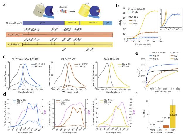 The fluorescent glutamate indicator iGluSnFR enables imaging of neurotransmission with genetic and
molecular specificity. However, existing iGluSnFR variants exhibit saturating activation kinetics and are
excluded from post-synaptic densities, limiting their ability to distinguish synaptic from extrasynaptic
glutamate. Using a multi-assay screen in bacteria, soluble protein, and cultured neurons, we generated novel
variants with improved kinetics and signal-to-noise ratios. We also developed surface display constructs that
improve iGluSnFR’s nanoscopic localization to post-synapses. The resulting indicator, iGluSnFR3, exhibits
rapid non-saturating activation kinetics and reports synaptic glutamate release with improved linearity and
increased specificity versus extrasynaptic signals in cultured neurons. In mouse visual cortex, imaging of
iGluSnFR3 at individual boutons reported single electrophysiologically-observed action potentials with high
specificity versus non-synaptic transients. In vibrissal sensory cortex Layer 4, we used iGluSnFR3 to
characterize distinct patterns of touch-evoked feedforward input from thalamocortical boutons and both
feedforward and recurrent input onto L4 cortical neuron dendritic spines. The fluorescent glutamate indicator iGluSnFR enables imaging of neurotransmission with genetic and
molecular specificity. However, existing iGluSnFR variants exhibit saturating activation kinetics and are
excluded from post-synaptic densities, limiting their ability to distinguish synaptic from extrasynaptic
glutamate. Using a multi-assay screen in bacteria, soluble protein, and cultured neurons, we generated novel
variants with improved kinetics and signal-to-noise ratios. We also developed surface display constructs that
improve iGluSnFR’s nanoscopic localization to post-synapses. The resulting indicator, iGluSnFR3, exhibits
rapid non-saturating activation kinetics and reports synaptic glutamate release with improved linearity and
increased specificity versus extrasynaptic signals in cultured neurons. In mouse visual cortex, imaging of
iGluSnFR3 at individual boutons reported single electrophysiologically-observed action potentials with high
specificity versus non-synaptic transients. In vibrissal sensory cortex Layer 4, we used iGluSnFR3 to
characterize distinct patterns of touch-evoked feedforward input from thalamocortical boutons and both
feedforward and recurrent input onto L4 cortical neuron dendritic spines.
|