 In vivo Ca2+ imaging of neuronal populations in deep cortical
layers has remained a major challenge, as the recording depth of
two-photon microscopy is limited because of the scattering and
absorption of photons in brain tissue. A possible strategy to increase
the imaging depth is the use of red-shifted fluorescent dyes,
as scattering of photons is reduced at long wavelengths. Here, we
tested the red-shifted fluorescent Ca2+ indicator Cal-590 for deep
tissue experiments in the mouse cortex in vivo. In experiments involving
bulk loading of neurons with the acetoxymethyl (AM) ester
version of Cal-590, combined two-photon imaging and cell-attached
recordings revealed that, despite the relatively low affinity of Cal-
590 for Ca2+ (Kd = 561 nM), single-action potential-evoked Ca2+
transients were discernable in most neurons with a good signalto-
noise ratio. Action potential-dependent Ca2+ transients were
recorded in neurons of all six layers of the cortex at depths of up
to −900 μm below the pial surface. We demonstrate that Cal-590 is
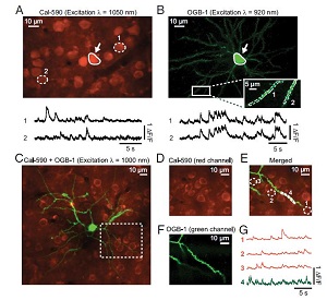
also suited for multicolor functional imaging experiments in combination
with other Ca2+ indicators. Ca2+ transients in the dendrites of
an individual Oregon green 1,2-bis(o-aminophenoxy)ethane-N,N,N′,
N′-tetraacetic acid-1 (OGB-1)-labeled neuron and the surrounding
population of Cal-590-labeled cells were recorded simultaneously
on two spectrally separated detection channels. We conclude that
the red-shifted Ca2+ indicator Cal-590 is well suited for in vivo twophoton
Ca2+ imaging experiments in all layers of mouse cortex. In
combinationwith spectrally different Ca2+ indicators, such as OGB-1,
Cal-590 can be readily used for simultaneous multicolor functional
imaging experiments.
In vivo Ca2+ imaging of neuronal populations in deep cortical
layers has remained a major challenge, as the recording depth of
two-photon microscopy is limited because of the scattering and
absorption of photons in brain tissue. A possible strategy to increase
the imaging depth is the use of red-shifted fluorescent dyes,
as scattering of photons is reduced at long wavelengths. Here, we
tested the red-shifted fluorescent Ca2+ indicator Cal-590 for deep
tissue experiments in the mouse cortex in vivo. In experiments involving
bulk loading of neurons with the acetoxymethyl (AM) ester
version of Cal-590, combined two-photon imaging and cell-attached
recordings revealed that, despite the relatively low affinity of Cal-
590 for Ca2+ (Kd = 561 nM), single-action potential-evoked Ca2+
transients were discernable in most neurons with a good signalto-
noise ratio. Action potential-dependent Ca2+ transients were
recorded in neurons of all six layers of the cortex at depths of up
to −900 μm below the pial surface. We demonstrate that Cal-590 is
also suited for multicolor functional imaging experiments in combination
with other Ca2+ indicators. Ca2+ transients in the dendrites of
an individual Oregon green 1,2-bis(o-aminophenoxy)ethane-N,N,N′,
N′-tetraacetic acid-1 (OGB-1)-labeled neuron and the surrounding
population of Cal-590-labeled cells were recorded simultaneously
on two spectrally separated detection channels. We conclude that
the red-shifted Ca2+ indicator Cal-590 is well suited for in vivo twophoton
Ca2+ imaging experiments in all layers of mouse cortex. In
combinationwith spectrally different Ca2+ indicators, such as OGB-1,
Cal-590 can be readily used for simultaneous multicolor functional
imaging experiments.