| Tracking stem cell differentiation in the setting of automated optogenetic stimulation
|
| Nov 18, 2010
|
| Stroh A, Tsai HC, Ping Wang L, Zhang F, Kressel J, Aravanis A, Santhanam N, Deisseroth K, Konnerth A, Schneider MB.
|
| Stem Cells
|
 Abstract
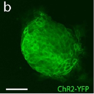
Membrane depolarization has been shown to play an important role in the neural differentiation of stem cells as well as in the survival and function of mature neurons. Here we introduce a microbial opsin into embryonic stem cells and develop optogenetic technology for stem cell engineering applications, with an automated system for noninvasive modulation of embryonic stem cell differentiation employing fast optogenetic control of ion flux. Mouse embryonic stem cells (ESCs) were stably transduced with ChR2-YFP and purified by FACS. Illumination of resulting ChR2-ESCs with pulses of blue light triggered inward currents. These labeled ESCs retained the capability to differentiate into functional mature neurons, assessed by the presence of voltage-gated sodium currents, action potentials, fast excitatory synaptic transmission, and expression of mature neuronal proteins and neuronal morphology. We designed and tested an apparatus for optically stimulating ChR2-ESCs during chronic neuronal differentiation, with high-speed optical switching on a custom robotic stage with environmental chamber for automated stimulation and imaging over days, with tracking for increased expression of neural and neuronal markers. These data point to potential uses of ChR2 technology for chronic and temporally precise noninvasive optical control of embryonic stem cells both in vitro and in vivo, ranging from noninvasive control of stem cell differentiation to causal assessment of the specific contribution of transplanted cells to tissue and network function. Abstract
Membrane depolarization has been shown to play an important role in the neural differentiation of stem cells as well as in the survival and function of mature neurons. Here we introduce a microbial opsin into embryonic stem cells and develop optogenetic technology for stem cell engineering applications, with an automated system for noninvasive modulation of embryonic stem cell differentiation employing fast optogenetic control of ion flux. Mouse embryonic stem cells (ESCs) were stably transduced with ChR2-YFP and purified by FACS. Illumination of resulting ChR2-ESCs with pulses of blue light triggered inward currents. These labeled ESCs retained the capability to differentiate into functional mature neurons, assessed by the presence of voltage-gated sodium currents, action potentials, fast excitatory synaptic transmission, and expression of mature neuronal proteins and neuronal morphology. We designed and tested an apparatus for optically stimulating ChR2-ESCs during chronic neuronal differentiation, with high-speed optical switching on a custom robotic stage with environmental chamber for automated stimulation and imaging over days, with tracking for increased expression of neural and neuronal markers. These data point to potential uses of ChR2 technology for chronic and temporally precise noninvasive optical control of embryonic stem cells both in vitro and in vivo, ranging from noninvasive control of stem cell differentiation to causal assessment of the specific contribution of transplanted cells to tissue and network function.
|