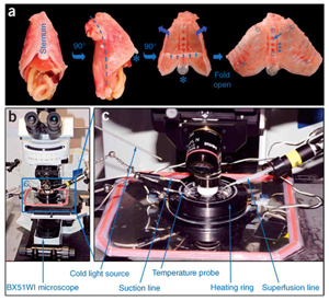
 We provide a protocol that describes an explant system that allows the dynamics of motor axons to be imaged. This method is based on nerve-muscle explants prepared from the triangularis sterni muscle of mice, a thin muscle that covers the inside of the thorax. These explants, which can be maintained alive for several hours, contain long stretches of peripheral motor axons including their terminal arborizations and neuromuscular junctions. Explants can be prepared from transgenic mouse lines that express fluorescent proteins in neurons or glial cells, which enables direct visualization of their cellular and subcellular morphology by fluorescence microscopy. Time-lapse imaging then provides a convenient and reliable approach to follow the dynamic behavior of motor axons, their surrounding glial cells and their intracellular organelles with high temporal and spatial resolution. Triangularis sterni explants can be prepared in 15 min, imaged ex vivo for several hours and processed for immunohistochemistry in about 2 h.